
fig 2.
Patient with a defect revealed by CT and a leak revealed by radionuclide cisternography but not by CT cisternography.
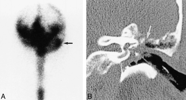
A, Radionuclide cisternogram of the head, anterior planar view, shows an abnormal accumulation of radionuclide (arrow) in the left mastoid region.
B, Coronal CT scan of the temporal bone shows a defect (arrow) in the tegmen tympani with adjacent mucosal thickening. The bone and dural defect were confirmed at surgery.