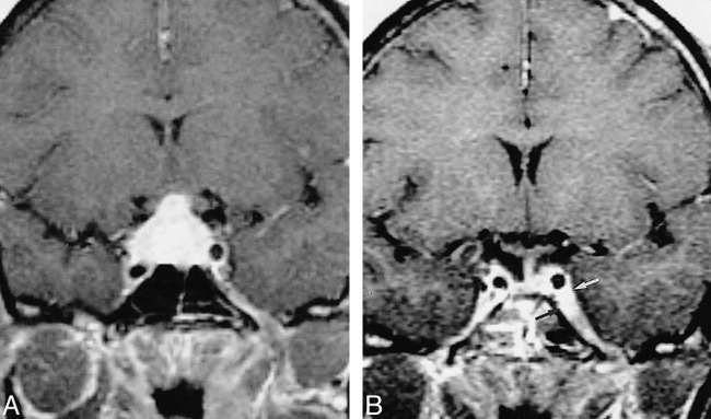
fig 2.

33-year-old African-American woman with right-sided visual loss, panhypopituitarism, polydipsia, and polyuria (with normal ADH).
A and B, T1-weighted contrast-enhanced MR image (400/15/2) (A) shows basal meningeal enhancement and an enhancing pituitary mass involving both lobes, the infundibulum, and the right optic nerve (not shown). Pituitary and meningeal biopsy revealed noncaseating granulomas with negative AFB and fungal stains. The visual symptoms resolved with steroid treatment; however, 21 months later she returned with a left abducens palsy. A T1-weighted image (400/12/2) shows resolution of the pituitary lesion but new involvement of the left cavernous sinus (arrows, B).
