
fig 3.
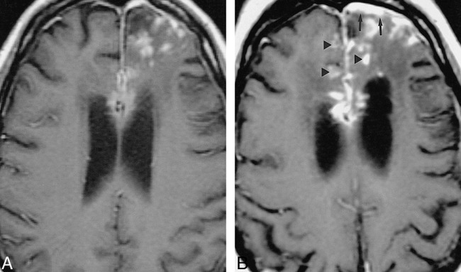
52-year-old African-American man with seizure disorder. Imaging revealed hydrocephalus and a brain parenchymal lesion. His seizures gradually became more difficult to control.
A and B, Contrast-enhanced T1-weighted MR images obtained 9 (483/13/2) (A) and 11 (500/16/2) (B) years after seizure onset show interim progression of meningeal thickening (arrows, B) and enlargement and multiplication of enhancing foci (arrowheads, B).