fig 1.

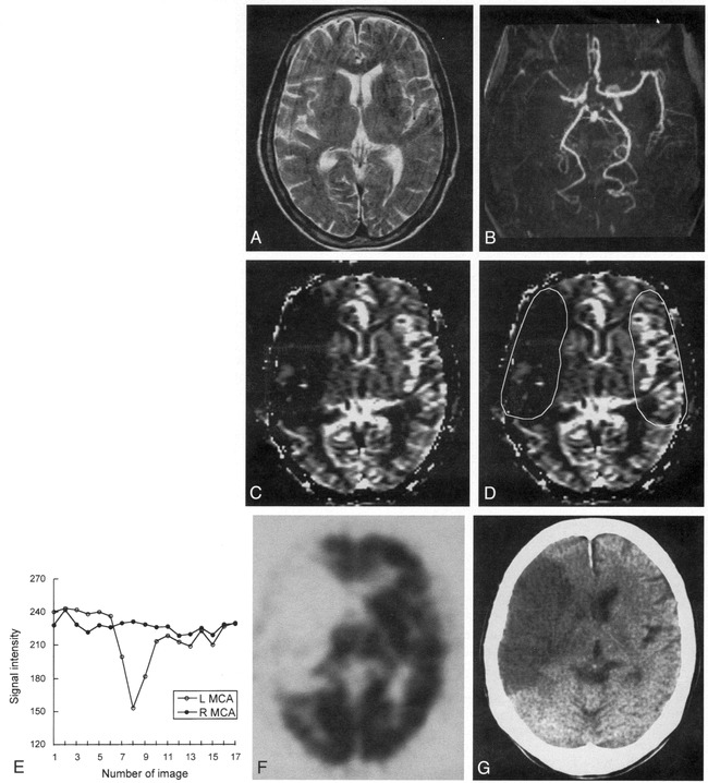
Case 8: 77-year-old woman with hyperacute ischemic stroke with arterial occlusion and decreased CBV.
A, T2-weighted MR image (3500/90) is normal except for abnormal subtle high signal intensity in right basal ganglia.
B, 3D time-of-flight MR angiogram reveals occlusion at M1 segment of the right MCA.
C, CBV map shows decreased CBV in right MCA distribution.
D, CBV map shows irregular ROIs placed for measurement of CBV ratio and time–signal intensity curves between the region of decreased CBV and contralateral normal region. Calculated CBV ratio was 0.17.
E, Time–signal intensity curves measured during passage of contrast material show no signal change in right MCA distribution compared with normal signal reduction in left MCA distribution.
F, 99mTc-HMPAO brain SPECT scan obtained during the hyperacute stage at approximately the same level as A and C reveals severe hypoperfusion throughout right MCA distribution.
G, Follow-up CT scan 3 days after the onset of symptoms shows well-defined infarction in right MCA distribution, which corresponds to the region of decreased CBV.
