fig 2.

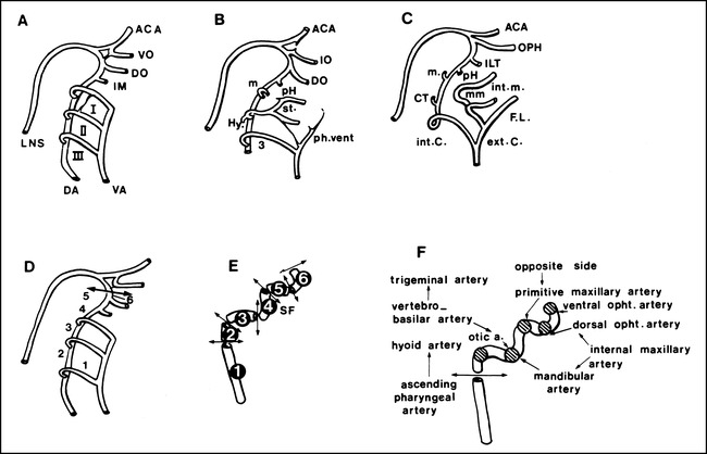
An illustration of the embryonic development of the aortic arches in the craniocephalic region is depicted. Successive stages of development are shown (A, B, and C), while the specific development of the internal carotid artery (D), its different segments (E) and the embryonic vessels and their origins (F) are highlighted.
A, Aortic arches (I—III) are numbered in the craniocaudal direction: the anterior cerebral artery (ACA); ventral ophthalmic artery (VO); dorsal ophthalmic artery (DO); primitive maxillary artery (IM); longitudinal neural system (LNS); dorsal aorta (DA), and ventral aorta (VA) are shown.
B, The primitive ophthalmic (IO), stapedial (st), hyoid (Hy), and ventral pharyngeal artery (vent. Ph.) arteries are illustrated.
C, The definitive ophthalmic artery (OPH), intracavernous collateral of the internal carotid artery (ILT) middle meningeal arteries (mm), caroticotympanic artery (CT), internal maxillary artery (int. m.), faciolingual system (FL), internal carotid (int. C) and external carotid artery (ext. C) are highlighted.
D, The third aortic arch (1), dorsal aorta between the second and third aortic arches (2), dorsal aorta between the first and second aortic arches (3), dorsal aorta between the first aortic arch and the primitive maxillary artery (4), dorsal aorta between the primitive maxillary artery and the inferolateral trunk (5), and dorsal aorta between the inferolateral trunk and its terminal branches (6) are shown.
E, The cervical segment (1), initial ascending intrapetrous segment (2), distal horizontal intrapetrous segment (3), segment ascending in the sphenoid fissure (SF) and through the cavernous sinus (4), horizontal segment of the carotid siphon (5), and clinoid segment (6) are shown.
F, Embryonic vessels and their origin are illustrated.
