fig 1.

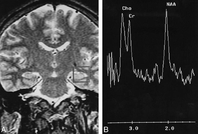
Proton SVS study of the left hippocampus of a healthy adult.
A, This coronal T2-weighted image (3000/85/1) is the central section for voxel positioning, about 1 to 2 mm anterior to the ipsilateral internal acoustic canal. The voxel is 20 × 20 × 20 mm3. Part of the hippocampal head and body are included for measurement.
B, There are three major peaks in this proton spectrum: NAA at 2.0 ppm, Cr at 3.0 ppm, and Cho at 3.2 ppm. The NAA peak is usually, but not necessarily, higher than the other two.
