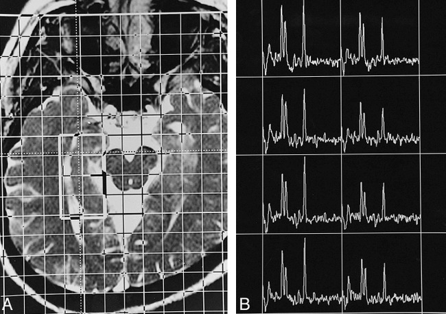
fig 2.

Proton CSI study of the right hippocampus of a healthy adult.
A, This transverse T2-weighted MR image (6.32/3.00/2), approximately parallel to the long axis of the hippocampus, illustrates the position of the VOI (about 40 × 20 mm2) inside the field of view (160 × 160 mm2), which has 16 × 16 phase encoding. The section thickness is 15 mm, the in-plane resolution is 10 × 10 mm2, and the voxel size is 10 × 10 × 15 mm3. The lateral margin of the VOI is about 0 to 2 mm lateral to the lateral border of the ipsilateral temporal horn, and the anterior margin is 5 to 10 mm anterior to the peduncles.
B, The resultant spectral map has eight voxels arranged in a two by four matrix. The spectral profile is similar to that of a single-voxel study. Owing to field inhomogeneity caused by anatomic complexity, in many cases the spectral resolution is worse in the more anterior voxels than in the posterior ones.
