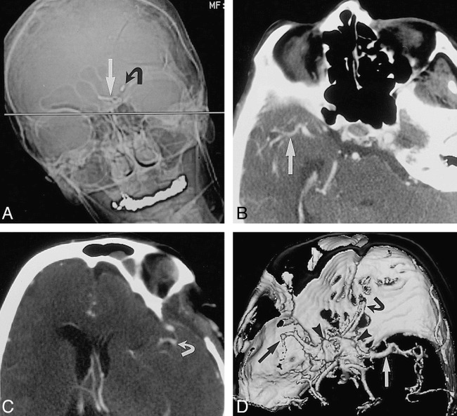
fig 1.

Tilted head position for intracranial CT angiography in a patient who has undergone clipping of multiple aneurysms.
A, Anteroposterior scout image of the head for planning CT angiography shows plane (straight line) for axial image acquisition (straight arrow indicates aneurysm clip in anterior communicating artery; curved arrow, aneurysm clip in carotid bifurcation). Note position of head should minimize image degradation, because metal artifact will project away from vessels of interest.
B and C, Axial source images from CT angiogram show right middle cerebral artery (arrow, B) and left middle cerebral artery (arrow, C) without interference from metallic beam-hardening artifact.
D, Shaded-surface-display image from CT angiogram viewed from above shows left (white arrow) and right (black arrow) middle cerebral artery without interference from aneurysm clip's beam-hardening artifact (arrowheads). Owing to limited z-axis coverage, left middle cerebral artery is abbreviated because examination was targeted to suspicious right middle cerebral artery. Current CT scanner software would allow coverage of entire head, tilted or not.
