
fig 2.
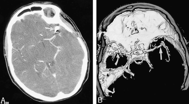
Tilted head position for intracranial CT angiography in a patient who has undergone clipping of a single aneurysm.
A, Axial source image from CT angiogram shows left middle cerebral artery aneurysm clipping (arrow) with adjacent metallic beam-hardening artifact.
B, Shaded-surface-display image from CT angiogram viewed from above shows right middle cerebral artery (straight arrow) without interference from aneurysm clip's beam-hardening artifact (curved arrow).