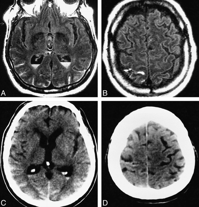
fig 1.

Case 12: 70-year-old man with subacute aneurysmal IVH and SAH who had MR imaging 9 days after ictus and CT 8 days after ictus. The FLAIR images show each occurrence of hemorrhage more conspicuously than do the CT scans. Although both FLAIR MR imaging and CT depicted IVH, the FLAIR images more crisply show the fluid-fluid levels characteristic of IVH.
A and B, FLAIR images (6700/150/2, TI = 2200) show layered IVH in the trigones of the lateral ventricles, which are hyperintense (A), resulting in blood-CSF fluid-fluid levels. Mixed intensities in the third ventricle are most likely caused by CSF pulsation artifact (and were also seen on the control images). Hyperintense posterior bilateral parietal and occipital (A) and right rolandic (B) cortical sulci represent SAH.
C and D, Noncontrast CT scans show both the IVH and SAH less conspicuously than do the FLAIR images, including layered IVH (C), bilateral parietooccipital SAH (C), and right rolandic sulcus SAH (D).
