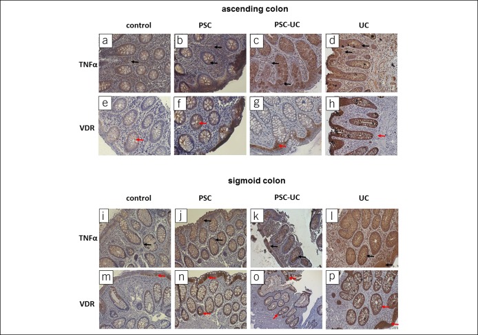
Figure 3.
Immunohistochemical localization of VDR and TNF-α proteins in human intestinal tissue. Representative immunostaining of colonic biopsies from controls, patients with PSC, patients with PSC-UC, and patients with UC with anti-TNF-α antibodies (a–d and I–l) and anti-VDR antibodies (e–h and m–p). Brown staining indicates either VDR protein (red arrows), which is located typically in the epithelial cells, or TNF-α protein (black arrows), which is depicted in expanded apical portions of goblet cells. Nuclei were visualized by hematoxylin. Original magnification was 200×. PSC, primary sclerosing cholangitis; TNF-α, tumor necrosis factor-α; VDR, receptor of vitamin D.

