
fig 2.
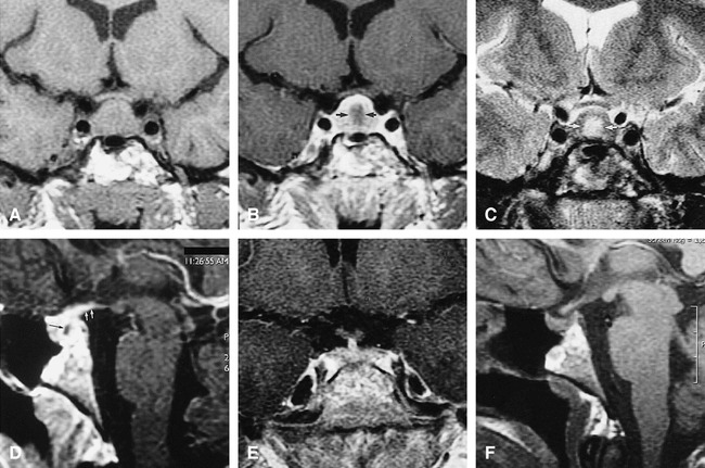
Case 2: MR findings in the sella turcica of 18-year-old woman with Wegener's granulomatosis.
A, Coronal T1-weighted (500/12/3) CSE image at initial presentation shows an enlarged pituitary gland (15 mm vertical height).
B, Coronal T1-weighted (500/12/3) CSE contrast-enhanced image shows enhancing periphery with relatively hypointense center (arrows).
C, Coronal T2-weighted (2433/95/4) CSE image with central hyperintensity (arrows) corresponding to the relatively hypointense region.
D, Sagittal T1-weighted (500/12/3) CSE contrast-enhanced image shows enhancing periphery with relatively hypointense center (black arrow) as well as enhancement into hypothalamus (white arrows).
E, Coronal T1-weighted (400/9/4) CSE contrast-enhanced image after long-term immunosuppressive therapy shows size of pituitary gland has decreased to normal range (7 mm in height) with normal, uniform enhancement.
F, Sagittal T1-weighted (400/9/4) CSE image after immunosuppressive therapy shows persistent loss of normal posterior pituitary hyperintensity.