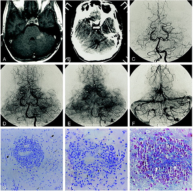
fig 1.

Patient 1.
A, Contrast-enhanced axial T1-weighted MR image (600/14/2) shows an enhancing lesion in the left brachium pontis.
B, Contrast-enhanced CT scan at the same level as the MR image. Although there is considerable streak artifact in the posterior fossa, there is an enhancing lesion in the left brachium pontis (arrow) corresponding to that identified on the MR image.
C–F, Early arterial (C), mid-arterial (D), capillary (E), and venous (F) phase images from the left vertebral artery stereo angiogram (anteroposterior projection) show an abnormal parenchymal blush (double arrows) in the left cerebellar hemisphere corresponding to the lesion seen on the CT and MR studies. There is arteriovenous shunting with early opacification of the left lateral recess and cerebellomedullary veins (curved arrows), which empty into the left sigmoidal sinus.
G, Histopathologic specimen shows exuberant perivascular inflammation (inf). The surrounding tissue shows gliosis (curved arrows) and microglial activation (straight arrows) (hematoxylin-eosin, original magnification ×100).
H, Higher-power view shows the mixed population of inflammatory cells, including lymphocytes, monocytes, and plasma cells (arrowheads). Note the plumping of the endothelial cells (arrow) (hematoxylin-eosin, original magnification ×200).
I, Azocarmine stain of tissue specimen in G shows reduplication of the basal lamina (arrows), seen as a fine connective tissue network (azocarmine stain, original magnification ×200).
