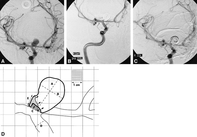
fig 2.

Coil compaction.
A, Pretreatment angiogram.
B, Posttreatment angiogram.
C, Follow-up angiogram at 7 months of a wide-necked (4.6 mm) carotid termination aneurysm.
D, Traced image (artist's rendition) derived from A, B, and C. The shaded area represents the residual uncoiled lumen at the time of treatment. The area filled by oblique lines is the additional lumen exposed by coil compaction. This is an example of initial treatment success (rest size < 2 mm) converting to failure at follow-up (rest size = 3.4 mm). For clarity, only the 1-cm calibrations have been rendered, with a portion of the figure showing the full 1-mm grid lines. “D” = linear reference diameter; “A” = luminal size (12.2 mm); “B” = luminal width (8.6 mm); “C” = neck size (4–6 mm); “E” = rest size at follow-up (3.4 mm).
