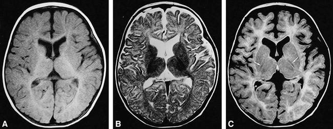
fig 4.

Brain MR images of a 30-week-old infant.
A, T1-weighted (500/15/2) image shows high signal intensity in the posterior limb of the internal capsule. The occipital white matter shows high intensity, suggesting myelination.
B, T2-weighted (4000/120/2) SE image shows low signal intensity of the posterior limb of the internal capsule.
C, FLAIR (8000/120/1, TI = 2000) image shows decreased signal intensity the posterior limb of the internal capsule (arrows). The occipital white matter shows increased signal intensity.
