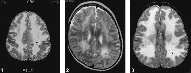
fig 1.

Patient 1: T2-weighted MR image of the brain at age 9 years shows diffuse and symmetrical increase in signal in the white matter of the cerebral hemispheres (arrow).
fig 2. Patient 2: T2-weighted MR image of the brain at age 8 years shows diffuse and symmetrical increase in signal in the white matter of the cerebral hemispheres (arrow).
fig 3. Patient 3: T2-weighted MR image of the brain at age 3 years shows diffuse increased signal in the white matter (arrow).
