
fig 2.
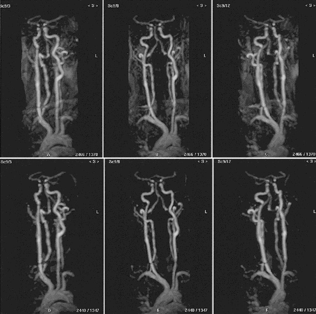
Rotated MIP projections of the eighth dynamic volume (arterial phase) acquired from a male volunteer using 3D dynamic keyhole turbo field-echo MR angiography (TR/TE = 6/3, flip angle = 50°). The nonsubtracted (top row) and the subtracted (bottom row) images show the aortic arch and major branches, the carotid arteries and bifurcations, and the vertebrobasilar arteries without interference from veins. Note the improved contrast between the arteries and background on the subtracted images