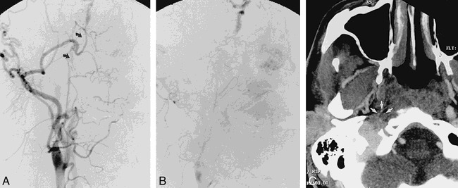
fig 4.

Frontal right carotid angiograms in 39-year-old man approximately 5 months after right carotid dissection with occlusion.
A and B, Short-segment serpiginous network of vessels bridge narrowed cervical ICA to petrous portion of ICA (arrows).
C, CT angiogram, transverse view, shows multiple small vessels (arrows) at perimeter of reduced-caliber thrombosed ICA.
