fig 1.

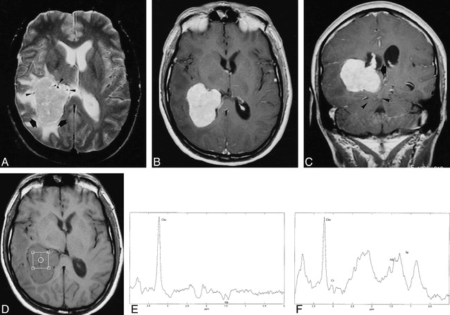
48-year-old man with a 2-month history of recurrent episodes of unsteady gait, visual loss, tinnitus, left arm paresthesia, and frontal headache.
A, Axial T2-weighted MR image shows an intraventricular meningioma in the right trigone (arrows) with heterogeneous signal, surrounded by edema. There are some enlarged vessels at the periphery of the mass (arrowheads).
B and C, Axial (B) and coronal (C) T1-weighted contrast-enhanced MR images show intense relatively homogeneous enhancement of the meningioma. Note contact with the choroid plexus (arrow, C) and posterior transtentorial herniation (arrowheads, C).
D, Axial T1-weighted MR image shows the position of the voxel for spectroscopy.
E, Localized proton MR spectrum (SE 2000/136/128) of the intraventricular meningioma shows a prominent resonance from Cho and a doublet centered at 1.47 ppm and inverted at this TE, which can be assigned to Ala.
F, Localized proton MR spectrum (SE 2000/31/128) shows a prominent resonance from Cho, a decrease in the Cr and NAA resonances, and the presence of lipids. Ala can also be assigned at this TE.
