
fig 3.
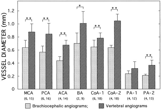
Vessel diameters, with and without brachiocephalic occlusion, were measured on brachiocephalic angiograms without occlusion (three images in three dogs) and vertebral angiograms with partial brachiocephalic occlusion (nine images in three dogs). The numbers of measured vessels on both sides are indicated in parentheses. The brachiocephalic angiogram of one representative dog is shown in figure 2. MCA indicates middle cerebral artery; PCA, posterior cerebral artery; ACA, anterior cerebellar artery; BA, basilar artery; CoA-1, communicating artery between MCA and PCA; CoA-2, CoA between ACA and BA; PA-1 and PA-2 are the anterior and posterior vessels of the two small branches of the circle of Willis, respectively. Data are means ± SD. *P = .053, **P < .001 (unpaired t-test)