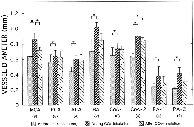
fig 5.

Effects of CO2 inhalation on changes in vessel diameters in the brains. Vessel diameters were measured on brachiocephalic angiograms before CO2 inhalation, during CO2 inhalation (2 L/min), and 60 minutes after completion of CO2 inhalation in three dogs. The numbers of vessels examined on both sides are indicated in parentheses. Images from one representative dog are shown in figure 4. In one dog, the basilar artery was outside the field of view. Abbreviations are the same as in figure 3. Data are means ± SD. *P < .05 (ANOVA)
