fig 1.

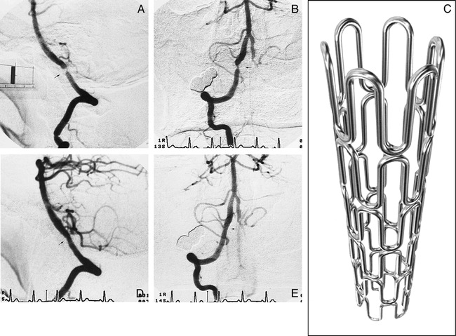
72-year-old man with an unruptured aneurysm in the right middle cerebral artery and total occlusion of the left vertebral artery.
A, Lateral view of the right vertebral arteriogram before stenting reveals a high-grade eccentric stenosis (arrow) with 93% stenosis and 4.5-mm length. Scale bar: 10 mm.
B, Anteroposterior view of the right vertebral arteriogram before stenting.
C, Illustration shows the gfx stainless steel stent, which was implanted in the intracranial vertebral artery. This stent measures 12 mm in length and can be expanded to up to 4 mm in diameter.
D and E, Right vertebral arteriograms (D, lateral view; E, anteroposterior view) after stenting show smooth and sufficient dilatation of the lesion and no residual stenosis (arrows).
