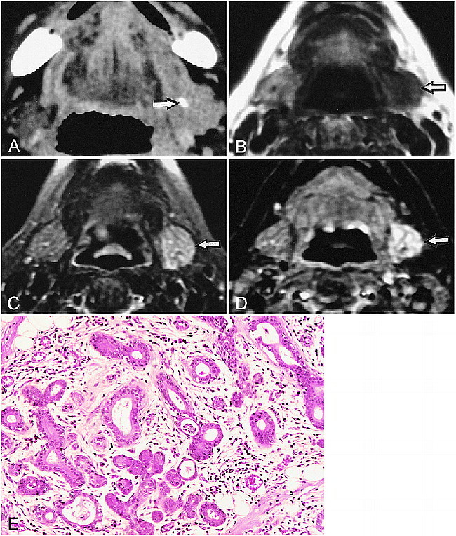
fig 1.

68-year-old woman with left submandibular sialolith.
A, CT shows sialolith (arrow) in hilum of the gland.
B, T1-weighted MR image (530/17/2 [TR/TE/excitations]) of affected submandibular gland (arrow) shows lower signal intensity compared with normal gland on right side.
C, Fat-suppressed T2-weighted MR image (3200/96/2 [TR/TE/excitations]) of affected submandibular gland (arrow) shows higher signal intensity than control gland (right side).
D, STIR image (2000/14/160/2 [TR/TE/inversion time/excitations]) of affected gland (arrow) shows higher signal intensity compared with normal gland on right side.
E, Photomicrograph of excised gland shows extensive infiltration of inflammatory mononuclear cells associated with destruction of gland structures and mild fibrosis. (Hematoxylin & eosin stain, original magnification, ×200)
