
fig 5.
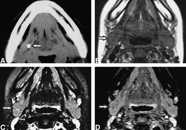
15-year-old girl with right submandibular sialolith.
A, CT shows sialolith (arrow) in proximal one third of main duct.
B, C, and D, T1-weighted (530/17/2 [TR/TE/excitations])(B), fat-suppressed T2-weighted (3200/96/2 [TR/TE/excitations]) (C), and STIR (2000/14/160/2 [TR/TE/inversion time/exciations])(D) MR images show no apparent difference between affected (arrow) and normal glands (left side).