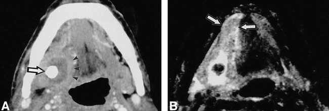
fig 6.

64-year-old man shows abscess formation in the extraglandular space.
A, Enhanced CT shows abscess formation around a large sialolith (arrow) in right floor of the mouth, demarcated by a slightly enhanced, broad rim (arrow heads). It was confirmed at surgery thas this sialolith was penetrating the duct wall.
B, T2-weighted MR image (3000/96/2 [TR/TE/excitations]) shows extension of inflammation in the floor of the mouth, beyond affected gland involving right sublingual gland (arrow).
