fig 1.

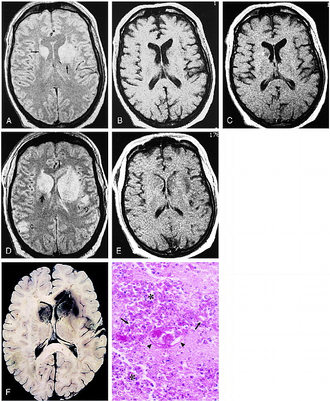
48-year-old man, 3 weeks after liver transplantation, with decline in mental status and flaccidity in bilateral lower extremities and left upper extremity.
A–C, Axial proton density–weighted (3000/25/2 [TR/TE/excitations]) (A) and unenhanced (500/20/2) (B) and contrast-enhanced (530/20/2) (C) T1-weighted spin-echo images. Areas of hyperintensity are present in the basal nuclei bilaterally, as well as in the left internal capsule on the proton density–weighted image (arrows, A), with corresponding modest hypointensity on the T1-weighted images. A small focus of enhancement is seen in the right caudate head (arrow, C).
D and E, Axial proton density–weighted (3000/25/2) and contrast-enhanced T1-weighted (530/20/2) spin-echo images obtained 6 days later show new involvement of the right internal capsule (large solid arrow) and callosal genu (small solid arrow). Cortical and subcortical lesions are also now seen (open arrows). Although the lateral ventricles are now effaced, there is rather little enhancement. The patient died several hours after this study was obtained.
F, The horizontally sectioned gross specimen shows red-brown areas of hemorrhagic necrosis corresponding to the abnormalities seen in D in the basal nuclei, and in the cortex and subcortical white matter. Hemorrhage is also seen in the genu of the corpus callosum.
G, Histologic section shows extensive necrosis with infiltration by polymorphonuclear leukocytes and macrophages (asterisks). Numerous Aspergillus hyphae are scattered throughout the section (arrows), and vascular fibrinoid necrosis is present (arrowheads) (PAS, hematoxylin; original magnification ×40).
