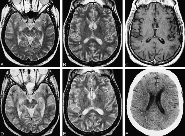
fig 3.

51-year-old man after bone marrow transplantation for IgA multiple myeloma with mental status decline. Pulmonary aspergillosis had been proved by bronchoalveolar lavage. A–C were obtained at neurologic presentation, D–F 5 months later.
A, Axial fast spin-echo T2-weighted (2200/84eff /1) image shows a mesencephalic lesion centered in the left substantia nigra (arrow).
B, Axial fast spin-echo T2-weighted (2200/84eff /1) image shows a hyperintense white matter lesion in the right parietal lobe (arrow). This patient also had severe (proved) bacterial sinusitis.
C, Axial fast spin-echo T1-weighted (600/12/2) image with contrast shows no definite enhancement of the lesion (arrow).
D, Axial fast spin-echo T2-weighted (2200/84eff /1) image of the midbrain shows return to normal after treatment with amphotericin B lipid complex.
E, Axial fast spin-echo T2-weighted (2200/84eff /1) image shows marked central hypointensity at the site of the parietal white matter lesion (arrow). There was again no enhancement of this lesion (image not shown).
F, Axial unenhanced CT scan shows high attenuation at the site of the parietal white matter lesion (arrow), confirming calcification of this lesion.
