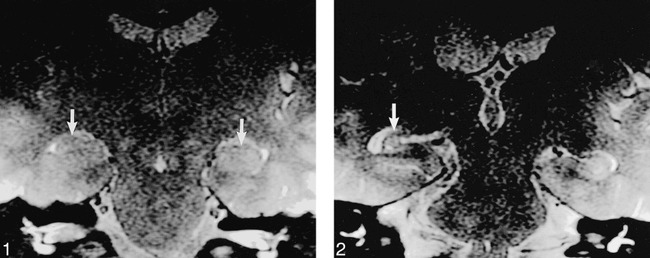
fig 1.

19-year-old nonepileptic patient with hearing loss. T2-weighted FSE (4000/80/3) image shows normal hippocampi bilaterally (arrows). Incidentally noted is a right acoustic schwannoma.
fig 2. Case 1: 29-year-old woman with new seizure onset 17 months after AVM hemorrhage. T2-weighted image (4000/80/3) obtained using the screening ear protocol 17 months after initial AVM hemorrhage shows unilateral right hippocampal sclerosis (arrow) with ipsilateral temporal lobe volume loss. No confirmatory high-resolution temporal lobe imaging was performed because the patient was not considered to be a surgical candidate.
