fig 1.

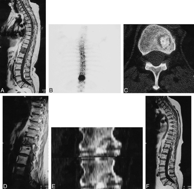
A 68-year-old woman with acute back pain of 15-day duration. Schmörl's node formation with herniation of calcified disk.
A, T1-weighted image spin-echo (600/15 [TR/TE]) shows diffuse low signal intensity in the T11 vertebral body and spondylolisthesis of L5–S1. T2-weighted spin-echo image (2200/80) shows only slight increase in signal intensity near inferior endplate (arrow).
B, Increased uptake on bone scintigraphy over T11 vertebra 6 days later.
C, CT scan depicts a radiolucent lesion in lower portion of T11 with central dense bone 14 days later.
D, Small defect (arrow) in inferior vertebral endplate of T11 and decrease in vertebral hypointensity on T1-weighted spin-echo image (500/15) 3 months later.
E, CT coronal reformatted images. Disk calcification herniating through vertebral bony defect.
F, T1-weighted spin-echo image (460/17) and T2-weighted spin-echo image (5000/112) (arrow) show a typical Schmörl's node at 1-year follow-up.
