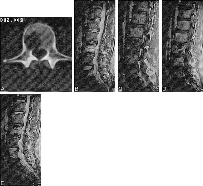
fig 2.

A 38-year-old man with 6-week history of lumbar pain related to physical exertion. Schmörl's node formation on neoplastic vertebra.
A, CT shows lytic lesion in L3 vertebral body without sclerotic margin.
B, T2-weighted spin-echo image (5000/90) shows hyperintense lesion in L3. Nucleus pulposus intranuclear clefts on L2–L3 and L3–L4 disks bend toward vertebral endplate.
C, T1-weighted spin-echo image (450/15) shows vertebral hypointensity and minimal irregularity on vertebral endplates.
D, Enhanced T1-weighted spin-echo image (450/15) shows two nonenhanced Schmörl's nodes of both endplates.
E, T2-weighted spin-echo image (5000/90) 3 months later shows a less hyperintense vertebral infiltrating lesion that extends to epidural space (arrow) and contains two Schmörl's nodes.
