
fig 1.
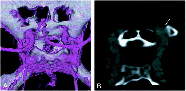
Patient with an infraclinoid, small aneurysm of the left internal carotid artery–ophthalmic artery.
A, 3D CT angiogram with SSD (superoposterior view) shows a slight laterally bulging contour (arrowhead); however, a significant saccular component cannot be identified.
B, Composite sagittal CPR image clearly shows a small aneurysm (arrow) adherent to the anterior clinoid process.