
fig 3.
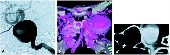
;t1Case 11: Patient with an aneurysm of the right internal carotid artery–ophthalmic artery.
A, Right carotid angiogram (left lateral view) shows a giant aneurysm.
B, 3D CT angiogram with SSD (superoposterior view) shows an aneurysm. The neck cannot be identified with the SSD technique owing to the giant overlapping fundus.
C, Composite sagittal CPR image clearly shows the neck of the aneurysm with the associated vessel of origin.