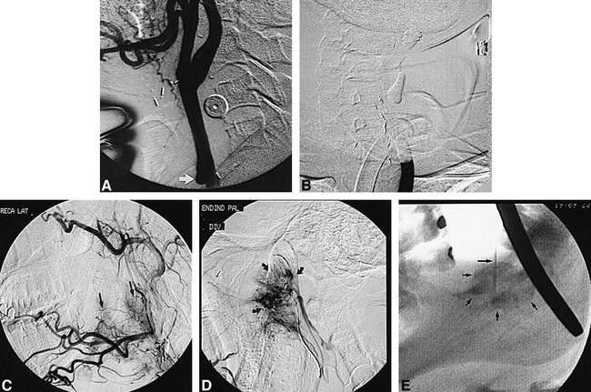
fig 1.

Illustrative case 1 of the spectrum of rCBS and its management.
A and B, Forty-six-year-old woman (patient TL) initially presents with CBS (Group 3). Lateral view from left common carotid injection (A) shows a pseudoaneurysm (arrow), which successfully was treated with therapeutic balloon occlusion (B).
C and D, Six years later, the patient develops a second episode of CBS. Lateral view from right external carotid injection (C) shows a hypervascular tumor of the oropharynx and hypopharynx (arrows) that is responsible for bleeding. Lateral view from superselective injection of the ascending palatine artery (D) shows significant supply to the tumor (arrows), which successfully was embolized with PVA.
E, Five days later, the patient develops a third episode of CBS, related to recurrent tumor hemorrhage. She was taken to the operating room in which, after induction of general anesthesia and oral retraction, the tumor was punctured under direct visualization and fluoroscopic guidance with a 23-gauge Chiba needle. Lateral fluoroscopic spot film shows the needle coursing through the tumor (long arrow) with extensive filling of neovasculature upon injection of absolute ethanol mixed with metrizamide (small arrows).
