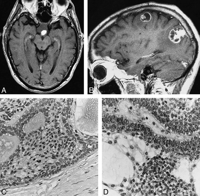
fig 1.

73-year-old man with metastatic craniopharyngioma.
A, In 1990, axial contrast-enhanced T1-weighted MR image (800/20 [TR/TE]) of the brain shows a 2-cm suprasellar mass with a solidly enhancing nodule along its posterior margin.
B, In 1997, left parasagittal contrast-enhanced T1-weighted MR image (540/14) of the brain shows two dural-based lobulated enhancing lesions. The left parietooccipital convexity lesion has a 2-cm cystic-appearing component attached to an enhancing dural-based pedicle. The left frontal lesion is smaller and has irregular mixed solid and rim enhancement characteristics.
C, In 1997, histologic section of the left parietooccipital lesion is composed of solid and cystic epithelial nests with distinct peripheral palisading and central loosely cohesive cells termed stellate reticulum (hematoxylin-eosin, original magnification ×400).
D, In 1990, the original suprasellar mass contains identical epithelial nests with peripheral palisading and central stellate reticulum (hematoxylin-eosin, original magnification ×400).
