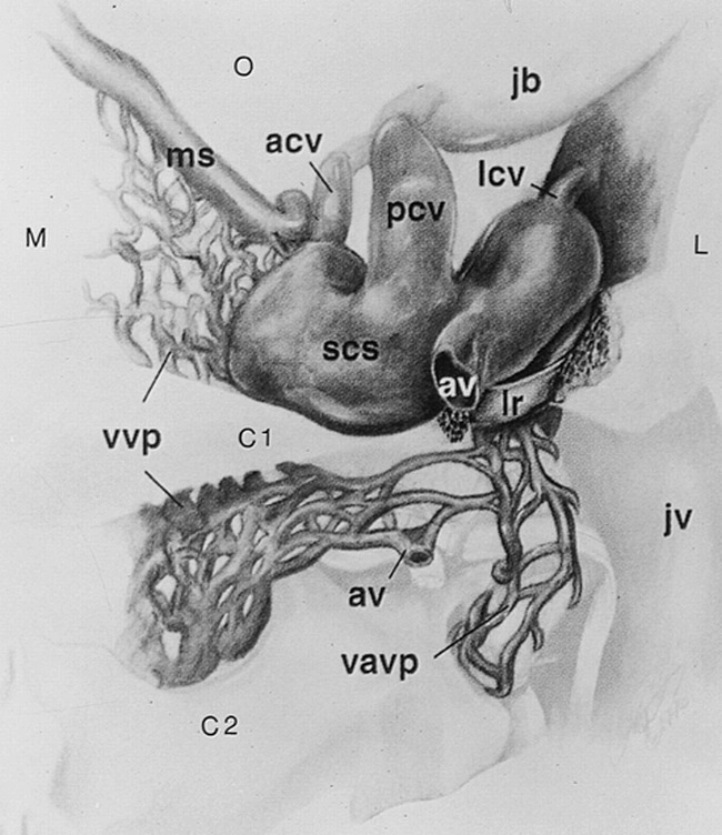
fig 1.

Posterior view of the right suboccipital cavernous sinus and its venous communications at the skull base. The vertebral artery has been removed. The left suboccipital cavernous sinus would form a mirror image, with direct connection of the marginal sinus and vertebral venous plexus across the midline. M indicates medial; L, lateral; C1, atlas; C2, axis; O, occipital region; acv, anterior condylar vein; av, anastomotic vein; jb, jugular bulb; jv, jugular vein; lcv, lateral condylar vein; lr, lateral ring; ms, marginal sinus; pcv, posterior condylar vein; scs, suboccipital cavernous sinus; vavp, vertebral artery venous plexus; vvp, vertebral venous plexus. Adapted from (1) with permission
