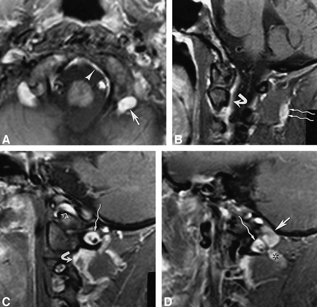
fig 5.

34-year-old woman.
A–D, Contrast-enhanced fat-suppressed T1-weighted (820/12/2) axial image (in a plane between fig 4A and B) (A) and T1-weighted (700/12/2) sagittal images (medial to lateral) (B–D) show the posterior condylar vein (straight arrow, A and D) at the posterior aspect of the occipital condyle, marginal sinus (arrowhead, A), suboccipital cavernous sinus (wavy arrow, C and D), vertebral venous plexus (curved arrow, B and C), suboccipital venous plexus (wavy arrows, B), anterior condylar vein (open arrow, C), and anastomotic vein (asterisk, D).
