
fig 2.
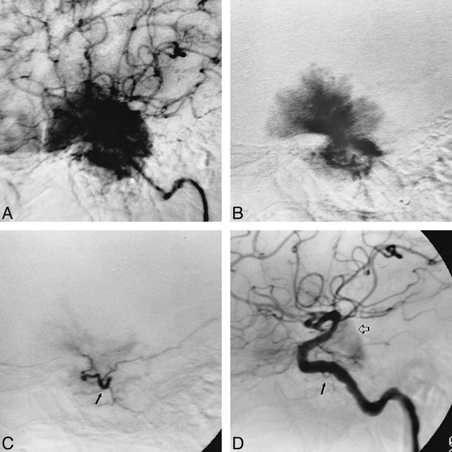
Case 3: 48-year-old woman with a left sphenoid wing meningioma.
A, Left ICA injection in mid-arterial phase shows an enlarged ILT branch and the early blush from the sphenoid wing meningioma.
B, Late arterial phase shows a dense tumor stain, which persisted late into the venous phase.
C, Selective injection of left ILT after direct catheterization with an extended tip Tracker-18 catheter. Capillary phase confirms supply to the meningioma via the enlarged ILT (arrow).
D, Left ILT injection after embolization with 2 mL of 150 to 250 μm PVA suspension. Late arterial phase shows abrupt cutoff of the posterior branch of the enlarged MHT (closed arrow).