
fig 3.
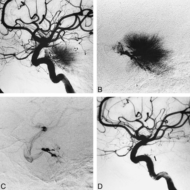
Case 5: 58-year-old woman with poor control of the left foot and left body hypesthesia.
A, Preembolization angiogram in late arterial phase during left ICA injection shows a dense tumor stain (open arrow) fed in part by left MHT branches (closed arrow).
B, Lateral view during selective injection in left MHT with an extended-tip, Tracker-18 catheter. Capillary phase confirms significant contribution to the tumor stain from left MHT.
C, Selective injection of left MHT after embolization with 2 mL of 150- to 250-μm PVA particle suspension shows near complete obliteration of the MHT supply to the tumor.
D, Left ICA injection after embolization confirms near complete devascularization of the tumor, with faint filling of the left tentorial marginal branch of the residual MHT (arrow).