
fig 4.
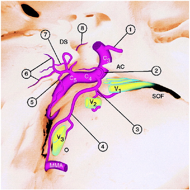
Diagram of the arteries arising from the cavernous segment of the ICA. AC, anterior clinoid; C3, C4, C5, segments of the intracavernous ICA; DS, dorsum sella; SOF, superior orbital fissure; O, foramen ovale; mma, middle meningeal artery; V1–V3, branches of the fifth cranial nerve coursing through superior orbital fissure (V1), foramen rotundum (V2), and foramen ovale (V3) accompanied by arterial branches of the ILT; 1, ophthalmic artery.
Branches of the ILT (C4 branches, 2 through 4):
2, Superior branch, supplying the roof of cavernous sinus; 3, anterior branch (to superior orbital fissure and foramen rotundum); 4, posterior branch (to foramen ovale and foramen spinosum).
Branches of the MHT (C5 branches, 5 through 8):
5, Recurrent artery of the foramen lacerum; 6, medial and lateral dorsal clival arteries; 7, tentorial marginal artery; 8, inferior hypophyseal artery.