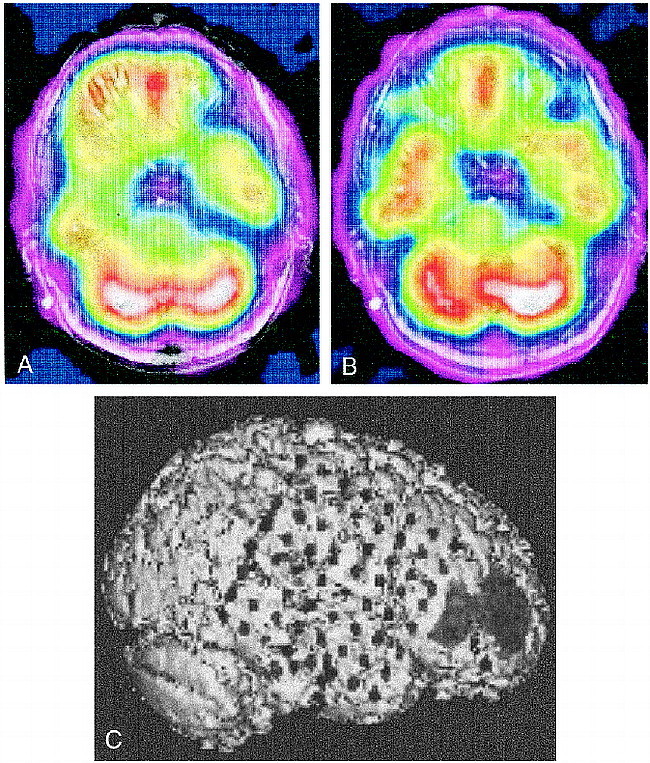
fig 3.

A, Interictal coregistered MR-SPECT scan with right temporal hypoperfusion.
B, Ictal coregistered MR-SPECT scan shows symmetrical bitemporal perfusion. This represents a relative increase in cerebral blood flow to the right temporal region during the seizure. There was ictal hyperperfusion in the right frontal region as well.
C, Triple-technique coregistration shows subdural strip electrodes over the right temporal and frontal regions in relation to the area of ictal hyperperfusion in the right frontal region. EEG recordings localized all seizures to the right temporal region.
