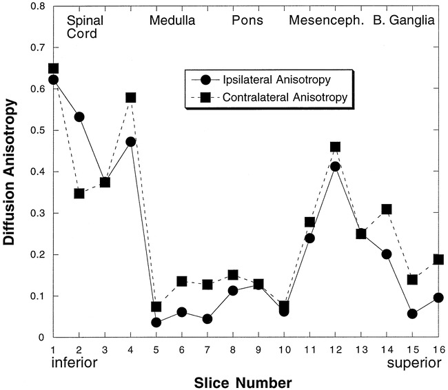
fig 2.

Diffusion anisotropy measurements following the corticospinal fiber tract in case 2. Measurements from 16 sections are shown. Sections are 5 mm thick with no gap between sections. Section 1 is the most inferior section. The diffusion anisotropy decreased bilaterally in the pons (sections 7–10) and parts of the medulla (sections 5 and 6) (see fig 3 for the locations of the axial sections)
