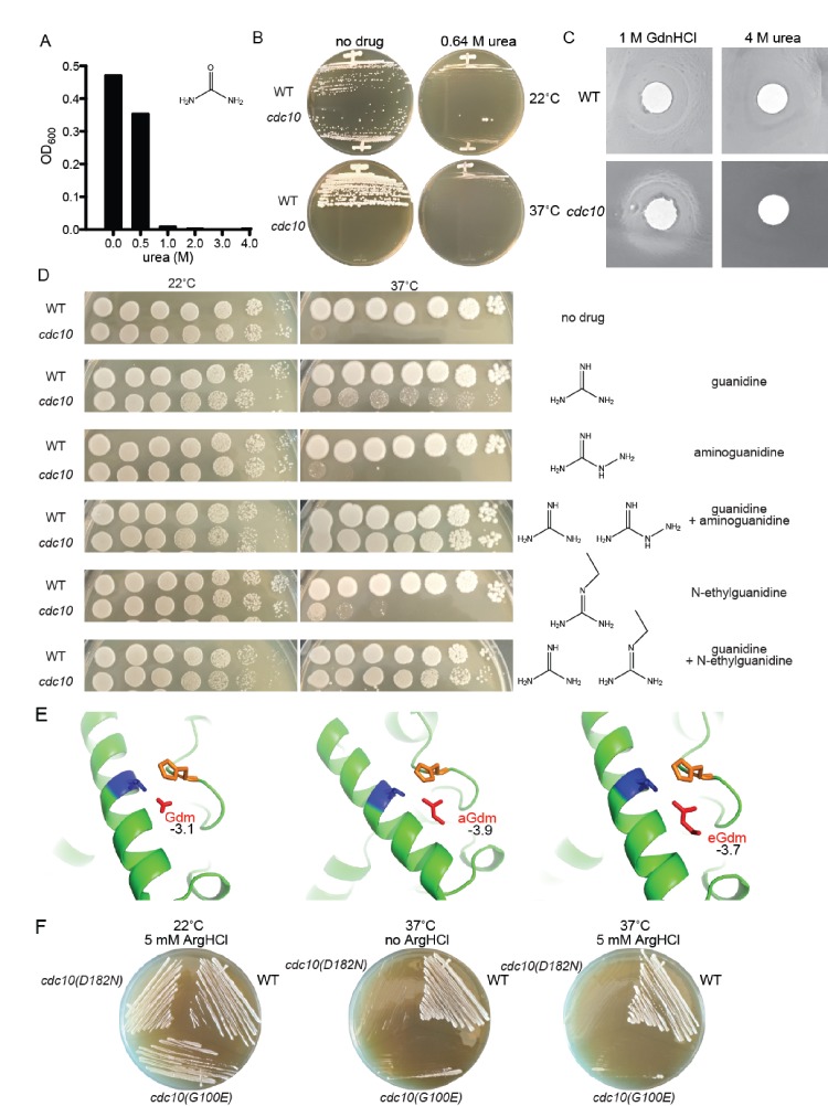
Figure 6. Partial rescue of TS defects by GdnHCl derivatives.
(A) WT strain BY4741 was cultured overnight in rich (YPD) medium containing the indicated concentrations of urea, and the density of each culture was determined with a spectrophotometer. (B) BY4741 (‘WT’) or the cdc10(D182N) strain CBY06417 (‘cdc10’) were streaked on plates containing no or 0.64 M urea and incubated at the indicated temperature for 3 days before imaging. (C) Cells from saturated YPD cultures of strains used in (A) were plated to form a monolayer on the surface of a rich (YPD) agar plate, and a 1-cm-diameter filter disk spotted with 5 µL of the indicated drug was placed in the center of the plate. The plate was imaged after incubation at 37°C for 3 days. (D) As in Figure 1A, for strains of the indicated genotypes grown on medium containing 0 or 0.375 mM of each indicated drug. (E) As in Figure 4A, but zoomed into the area between Thr302 and His262, and including the Gdm derivatives aminoguanidine (‘aGdm’) and N-ethylguanidine (‘eGdm’). (F) As in (B), but with the addition of strain CBY06420 (‘cdc10(G100E)') and with arginine hydrochloride instead of urea.

