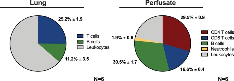
FIGURE 3.

Distribution of donor leukocytes in graft and perfusate compartments following EVLP. Three hours of EVLP was performed, followed by flow cytometric analysis of leukocytes isolated from the lung graft and the perfusate compartments. EVLP, ex vivo lung perfusion.
