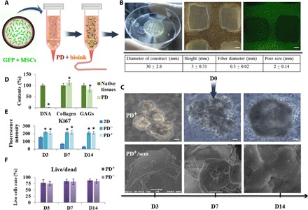
Fig. 1. Schematic illustration of 3D-bioprinted MSC-loaded constructs and cellular survival, proliferation, and morphology of printed construct.

(A) Schematic description of the approach. (B) Full view of the cellular construct and representative microscopic and fluorescent images and the quantitative parameters of 3D-printed construct (scale bars, 200 μm). Photo credit: Bin Yao, Wound Healing and Cell Biology Laboratory, Institute of Basic Medical Sciences, General Hospital of PLA. (C) Representative microscopy images of cell aggregates and tissue morphology at 3, 7, and 14 days of culture (scale bars, 50 μm) and scanning electron microscopy (sem) images of 3D structure (scale bars, 20 μm). PD+/PD−, 3D construct with and without PD. (D) DNA contents, collagen, and GAGs of native tissue and PD. (E) Proliferating cells were detected through Ki67 stain at 3, 7, and 14 days of culture. (F) Live/dead assay show cell viability at days 3, 7, and 14. *P < 0.05.
