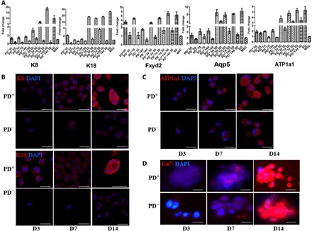
Fig. 2. Transcriptional and translational level of SG-specific and secretion-related markers in 3D-bioprinted cells with or without PD.

(A) Transcriptional expression of K8, K18, Fxyd2, Aqp5, and ATP1a1 in 3D-bioprinted cells with and without PD in days 3, 7, and 14 culture by quantitative real-time polymerase chain reaction (qRT-PCR). Data are means ± SEM. (B) Comparison of SG-specific markers K8 and K18 in 3D-bioprinted cells with and without PD (K8 and K18, red; DAPI, blue; scale bars, 50 μm). (C and D) Comparison of SG secretion-related markers ATP1a1 (C) and Ca2+ (D) in 3D-bioprinted cells with and without PD [ATP1a1 and Ca2+, red; 4′,6-diamidino-2-phenylindole (DAPI), blue; scale bars, 50 μm].
