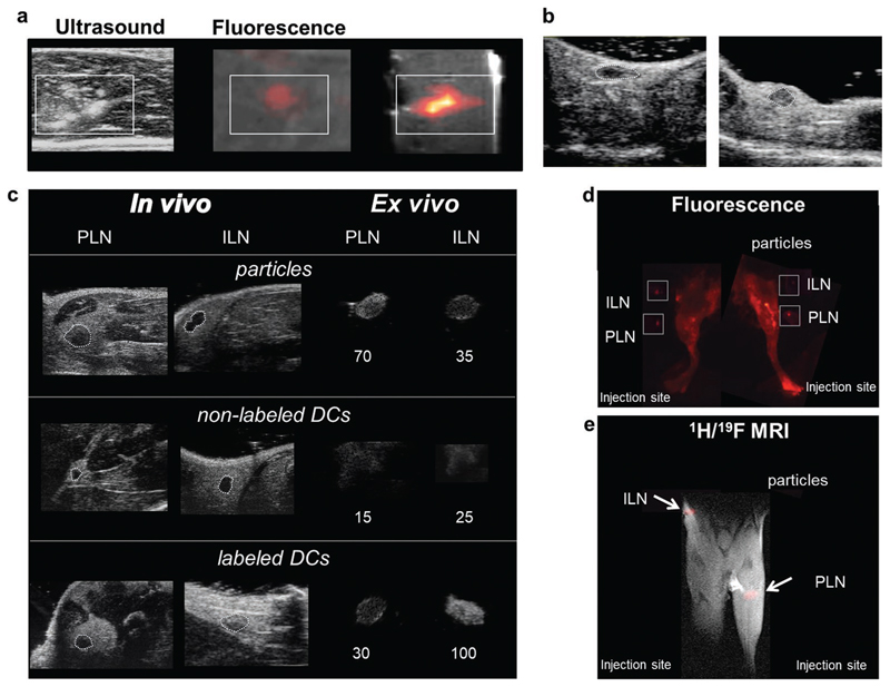
Figure 6.
Nanoparticles are suitable for tracking the therapeutic cells with ultrasound in vivo. a) US, fluorescence, and 1H and 19F (false color) MR images of 2 million DCs labeled with particles containing PFCE, IC-Green, and Gd injected in a tissue sample (boxes indicate position of the cells). b) High-frequency in vivo US images of the inguinal lymph node (ILN) of a mouse before (left) and after (right) intranodal injection of 0.1 mg of PFCE nanoparticles show a tenfold increase in mean contrast in the node after injection (see Videos S1 and S2, Supporting Information). c) Mice were injected with 5 million labeled primary murine DCs in the footpad and imaged after 24 h using US on a clinical scanner (Figure S9, Supporting Information) and a high-resolution small animal scanner (48 MHz). Control mice received an equivalent number of nonlabeled cells or free particles. In vivo images of the draining lymph nodes (inguinal lymph node (ILN) and popliteal lymph node (PLN)) are shown at high resolution (48 MHz). Ex vivo images are also shown (circled) at high and low frequency (Figure S9, Supporting Information). Labeled DCs increased contrast in the node nearly fivefold compared to nonlabeled cells. d) A fluorescence image shows the same results, with labeled cells mainly in the INL and particles in the PLN and surrounding lymphatics. Excised lymph nodes were also imaged (boxes). e) The corresponding 1H/19F MRI, with 19F data in false color, also shows the same distribution. The injection site is just outside the field of view. Please see Figure S11 (Supporting Information) for further in vivo images, and Figure S12 (Supporting Information) for ex vivo images on a clinical scanner.

