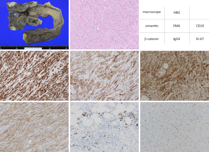
Figure 4.
Hematoxylin and Eosin (H&E) staining and immunohistochemistry staining were performed for the rectal ulceration in the resected specimen. The proliferating spindle cells were positively stained with vimentin, SMA, CD10 and β-catenin. Focal infiltration of IgG4-positive plasma cells was observed. About 30% of proliferating spindle cells were positively stained with Ki-67 in the hot spot. Vasculitis was not observed.

