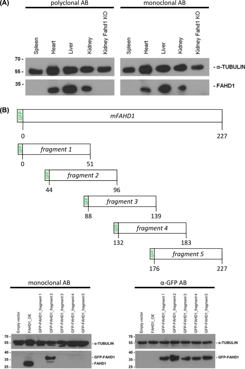
Figure 6. Identification of the epitope recognized by the monoclonal rabbit mFAHD1.
(A) Thirty microgram protein isolated from mouse tissue lysates were analyzed by Western blot, using a polyclonal α-mFAHD1 antibody and RabMab 27-1 (see ‘Materials and methods’ section). Kidney lysate of an FAHD1 knockout mouse was used as negative control, α-TUBULIN antibody (Sigma–Aldrich) was used as loading control. Lower molecular weight signal detected by the polyclonal antibody (not displayed) are non-specific and the intensity of the specific signal is roughly comparable between polyclonal and monoclonal antibodies. (B) Full-length FAHD1 was divided into five different constructs, encoding consecutive regions of the protein (aa 1–51; aa 44–95; aa 88–139; aa 132–183; aa 176–227) which were fused with GFP. Protein lysates from U2OS cells transfected with pcDNA_3.1_GFP-FAHD1 plasmids 1–5 were analyzed by Western blot analysis using mFAHD1 antibody and antibody directed against GFP. U2OS cells transfected with pcDNA_3.1_Hygro (-) (empty vector) served as negative control and cells transfected with Fahd1 overexpression plasmid (FAHD1_OE) as positive control.

