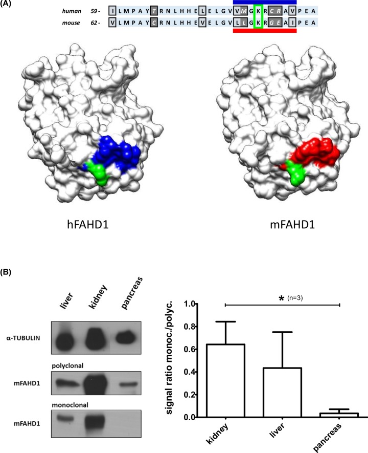
Figure 8. Lysine 83 in mFAHD1 is a reported SIRT3 deacetylation target.
(A) K83, K116, K117, and K142 of mFAHD1 have been reported to be deacetylation targets of SIRT3 [25]. K83 (K80 in hFAHD1) is in the center of the identified epitope region of the monoclonal antibody (Figure 7). The lysine side chain is depicted in green for both hFAHD1 and mFAHD1; residues comprising the putative epitope LLGKRGEAI in mouse FAHD1 are shown in red; the corresponding amino acid residues in hFAHD1 are depicted in blue. (B) Western blot of FAHD1 in an exemplary collection of mouse organs (wild-type and mFAHD1 knockout mice), similar to data presented elsewhere [23,24]. In contrast with the polyclonal antibody, RabMab 27-1 seems to be tissue specific, i.e., hindered in recognizing the antigen in certain tissues, such as pancreas (lowest plot, left subpanel). Western blot has been repeated three times (n=3), and the signals of polyclonal and monoclonal antibodies (normalized to the Tubulin signal) have been compared (right subpanel). Error bars represent the standard deviation within 95% confidence interval. We find that RabMab 27-1 recognized mFAHD1 in pancreas tissue, but the signals are significantly weaker than compared with kidney (*P < 0.05; P = 0.04).

