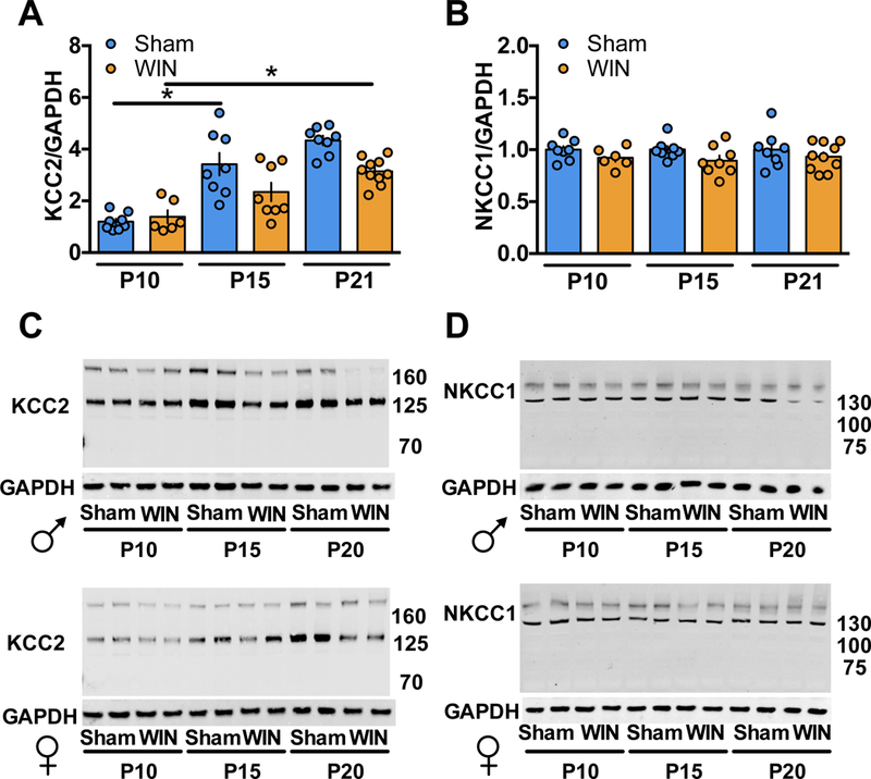
Figure 3: Perinatal WIN-exposure alters the developmental trajectory of KCC2 and NKCC1 expression in the mPFC.

Western-blot analysis of KCC2 and NKCC1 reveal altered expression levels between P10, P15 and P21 in progeny of dams exposed to WIN during lactation as compared to progeny of Sham-treated dams. A: KCC2 levels are significantly increased between P10 and P15 and remain elevated at P21 in the mPFC tissue collected from pups of Sham-treated dams (P10 N=8, P15 N=8, P21 N=8; F5,42=19.38, P<0.0001. One-way ANOVA followed by Tukey’s multiple comparisons test. Sham P10 vs. Sham P15, P<0.0001; Sham P15 vs. Sham P21, P=0.9744). However, no change in KCC2 levels was detected in mPFC tissue collected from pups of WIN-treated dams between P10 and P15 (P10 N=6, P15 N=8). At P21, a significant increase in KCC2 was observed in mPFC tissue from WIN-treated pups as compared to P10 (P21 N=10; P=0.0009, Tukey’s multiple comparisons test). B: No difference in NKCC1 levels was detected in mPFC tissue collected from pups of Sham- or WIN-treated dams at any of the tested time points (Sham P10 N=8, P15 N=8, P21 N=8; WIN P10 N=6, P15 N=8, P21 N=10). One-way ANOVA followed by Tukey’s multiple comparisons test, F5,42 = 1.108, p=0.3707. Error bars indicate SEM. *p<0.05. C,D: Representative Western-blots of KCC2/GAPDH and NKCC1/GAPDH, corresponding to a,b respectively.
